OCULAR DISEASE MANAGEMENT
Most eye conditions do not manifest symptoms until later stages. This is why we make it our priority to evaluate and detect ocular diseases while they are still preventable and treatable.
RETINAL COMPLICATIONS detection and treatment
During your eye exam, our doctors will evaluate the front surface as well as the inside of your eyes. We use an Optos retinal imaging system to document and detect ocular diseases.
This machine is used evaluate the retina in patients as young as 5 years old. Some conditions that can be detected with this cutting-edge device include macular degeneration, diabetic retinopathy, choroidal nevi, hypertensive retinopathy, retinal tears/holes/detachments, and retinal scarring. In addition to eye conditions, signs of other diseases such as heart disease, hypertension, and stroke risk can be detected as well. Dilation may also be used to further evaluate the retina.


CATARACT DETECTION and treatment
At your annual exam, our doctors will evaluate the lens inside your eye with a slit lamp to detect early stages of cataracts. A cataract is an opacity in the lens inside your eyes that can be caused by aging changes, UV damage, smoking, and diabetic complications. Small cataracts can be monitored and treated.
Since a cataract can cause glare issues and frequent shifts in one’s prescription, it is important to evaluate cataracts yearly to adjust your prescription and discuss options on glasses that can help you be more visually comfortable. If cataract surgery is needed, our doctors will make the appropriate referral and conduct the necessary follow-up evaluations to ensure the continuity of your care.
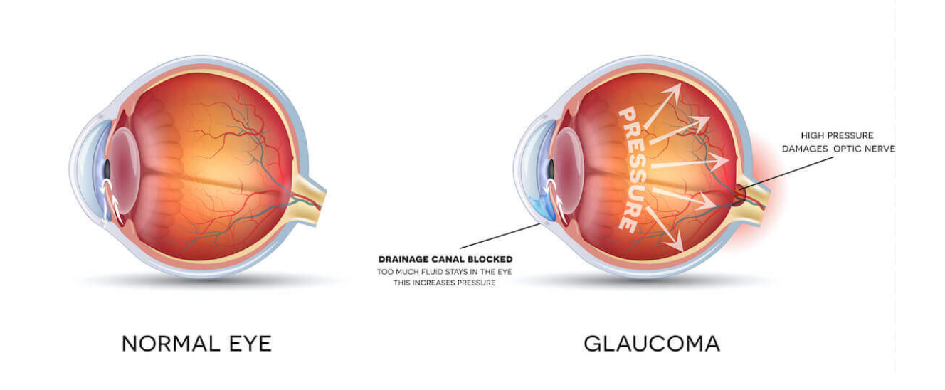
GLAUCOMA DETECTION and treatment
When you get your annual exam, our doctors will evaluate the pressure inside and check the optic nerve in your eyes. If further evaluation is needed, you may be asked to return for a glaucoma evaluation. The glaucoma evaluation often includes:
Optic nerve imaging documentation and interpretation
Virtual Field Visual Field Machine, which picks up loss in peripheral vision often before we are able to perceive these losses
Gonioscopy, which evaluates the drainage system in our eyes
The key in early glaucoma detection is to be proactive and have baseline information whenever there are any yellow flags at your regular exam. Once we have baseline information, we are better able to detect any structural changes in your eyes so that we can talk about effective treatment options that can help prevent further vision loss and blindness.
2.7 million people in the us have glaucoma
Glaucoma is a progressive condition that can potentially lead to permanent blindness. Glaucoma is often referred to as the Silent Stealer of Sight because most people don’t notice their vision loss until it is too late to treat and control.
Glaucoma is a very complex condition, with many variations as to the root cause. The most common form of glaucoma occurs when there is pressure built up in the eyes. The pressure can lead to damage to the optic nerve in the back of the eyes. If nerve signals stop sending visual information back to the brain, patients can eventually lose vision from the outside in (known as “tunnel vision”).